Please do not despair if your dog is diagnosed with mast cell tumors. We believe that through immunity-boosting efforts, you can improve your dog’s condition, maintain their quality of life (QOL), and restore their energy and appetite. In fact, there are numerous cases where cancer in dogs has been controlled through immune support using Cordy.
This page provides an overview of the characteristics of mast cell tumors, their treatment methods, and ways to improve prognosis. We also introduce many cases of improvement. We hope that this information can be a source of support and light of hope for you.
目次
- 1 What kind of cancer is mast cell tumor in dogs?
- 2 Symptoms when a dog has a mast cell tumor
- 3 Causes of Mast Cell Tumors
- 4 Tests When MCTs Are Suspected
- 5 Grading MCTs (Malignancy Level)
- 6 Treatment of Canine Mast Cell Tumors
- 7 Prevention Methods for Mast Cell Tumors in Dogs
- 8 Striving for Extended Life and Overcoming Mast Cell Tumors in Dogs
What kind of cancer is mast cell tumor in dogs?
What are mast cells?
Mast cells are named because they appear swollen and fat under a microscope.
Mast cells are a type of white blood cell that releases a substance called histamine. Histamine is involved in inflammation and immune responses and also affects the functioning of internal organs.
Although histamine may have a negative image due to its role in allergic reactions, such as causing symptoms in hay fever, it is actually a defense mechanism to expel foreign substances from the body. The body is protected by the function of mast cells.
It should be noted that adipocytes, which store fat within the cell, are related to obesity, but they have no direct connection with mast cells or mast cell tumors.
What is a mast cell tumor?
Mast cell tumors can occur in the skin or internal organs, but in dogs, they most commonly occur in the skin, making it the most common malignant tumor found in canine skin.
Although the name mast cell tumor might suggest a connection with obesity, there is no direct link between the two.

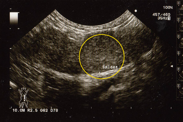
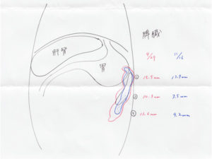
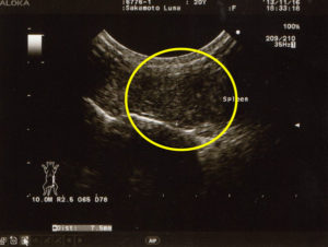
The image above is an ultrasound scan of a mast cell tumor. The drawing below is one used by veterinarians to explain to patients.

When a lump on the skin or internal organs is identified as a mast cell tumor, there is a high possibility it is malignant and therefore cancerous.
Due to the risk of spread or metastasis, it should be surgically removed promptly. However, because mast cell tumors have a high malignancy rate, a wide-margin excision, or ‘radical surgery‘ is necessary. Depending on the location, it might be challenging to secure adequate margins, and even if it appears to have been completely removed, recurrence or metastasis is not uncommon.
If surgery successfully removes the mast cell tumor without recurrence or metastasis, it is ideal. However, if it metastasizes and spreads to other organs or the entire body, it could cause issues such as gastrointestinal inflammation or bleeding, leading to blood in vomit or stools. Appetite loss and a decline in energy and appetite may also be observed.
Mast cell tumors are a type of malignant tumor, but early detection and prompt treatment can significantly improve the chances of recovery. Therefore, if you notice any abnormalities on your pet’s skin, please visit an animal hospital as soon as possible.
Symptoms when a dog has a mast cell tumor

Mast cell tumors in dogs can present in various shapes, ranging from hard lumps to soft fatty masses, or as nodules that either protrude like warts or spread flat across the skin. Due to this variation, it is necessary to undergo tests at an animal hospital to determine whether a lump is a mast cell tumor.
In mast cell tumors, substances like histamine are released in large quantities. While histamine is essential for life, excessive amounts can cause various issues such as increased susceptibility to allergic reactions, increased gastric acid leading to ulcers, and respiratory difficulties due to lung impairment. These issues can greatly reduce the quality of life (QOL) and, in severe cases, endanger life.
If pet owners notice changes in their dog’s appearance or health, they should visit an animal hospital as soon as possible and undergo necessary tests.
Symptoms when a mast cell tumor occurs on the skin
Most mast cell tumors in dogs develop on the skin. Approximately 50% occur on the trunk and genital area, 40% on the limbs, and 10% on the head and neck. Hair loss in these areas may also be an indicator.
When a lump develops on the dog’s skin and becomes inflamed, it might indicate a high degree of malignancy. If there is inflammation or bleeding from the lump, prompt examination by a veterinarian is recommended.
Other tumors found on the skin
Other types of skin cancer include melanoma and squamous cell carcinoma.
Symptoms when a mast cell tumor occurs in internal organs
Mast cell tumors can also be found in internal organs. Most cases in internal organs are considered to be metastases from the skin. Metastasis commonly occurs in lymph nodes, liver, spleen, and bone marrow.
When a mast cell tumor develops in internal organs, symptoms such as nausea and loss of appetite become prominent. There may be bloody stools due to internal bleeding, leading to anemia, and sometimes difficulty in defecation.
Causes of Mast Cell Tumors
Mast Cell Tumors (MCTs) can occur regardless of age or breed, and therefore, the exact cause of MCTs is still unknown.
Generally, genetic factors, a decline in immune function due to aging, lifestyle problems such as diet, environmental pollution, and stress are considered potential causes of MCTs.
MCTs tend to be more common in breeds such as Labrador Retrievers, Golden Retrievers, short-headed breeds like Pugs, Boxers, Bulldogs, but other breeds are also susceptible to MCTs.
Tests When MCTs Are Suspected
Cytology
Cytology involves using a needle to collect cells from the suspected MCT area and examining them under a microscope. This method is mostly painless and can be easily performed without anesthesia. However, as cytology yields a small number of cells, it sometimes fails to provide an accurate diagnosis.
Blood Tests
When MCTs are suspected, blood tests are conducted to check for anemia, assess internal organ function, and measure changes in histamine levels released by the MCTs.
Imaging Tests (X-ray, CT, MRI, etc.)
To check if the cancer has spread to internal organs or lymph nodes in areas such as the chest or abdomen, imaging tests like X-rays, CT scans, and MRI are performed. While imaging results alone cannot determine the type of tumor, a definitive diagnosis requires tissue biopsy or cytology.
Genetic Testing
Genetic testing, which involves collecting a small number of cells, can help determine the type of MCT. It is useful for predicting malignancy and the efficacy of certain treatments.
Grading MCTs (Malignancy Level)
All MCTs are malignant tumors (cancer), but they can be divided into three levels of malignancy, with treatment methods varying by severity. MCTs that develop outside the skin are treated as highly malignant.
| Grade 1 | This has a low malignancy level and is less likely to metastasize. Often, surgery alone is sufficient, and there are few recurrences. It offers the most promising chance for complete cure among MCTs. |
|---|---|
| Grade 2 | This has a higher malignancy level and may metastasize. Post-surgery, radiation therapy or chemotherapy may be administered. If recurrence or metastasis occurs, the prognosis is poor. |
| Grade 3 | This is extremely malignant and prone to metastasize. Even with surgery followed by chemotherapy or radiation, recurrence and metastasis occur frequently, making a complete cure difficult. |


